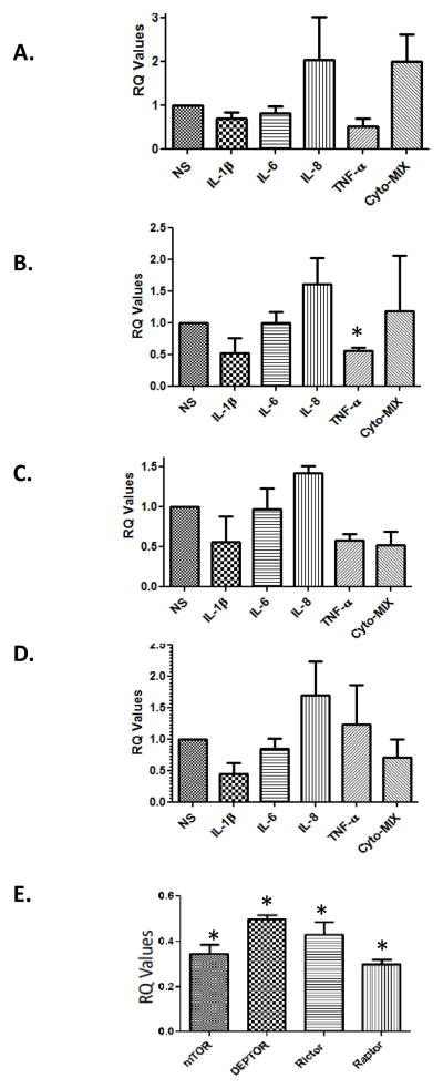
Figure 3. Progesterone but not IL-1β, IL-6, IL-8 down-regulated mTOR, DEPTOR, Rictor and Raptor, compared to unstimulated control cells.
Myometrial cells were treated with for 24 hrs with IL-1β, IL-6, IL-8 and TNF-α (1ng/mL) and levels of mTOR (A), DEPTOR (B), Rictor (C) and Raptor (D) were assessed. Only TNF-α significantly (*p<0.05) downregulated DEPTOR’s expression. Panel E: Cells were treated with 30nM P4 for 24 hrs and the levels of mTOR, DEPTOR, Rictor, and Raptor were assessed compared to basal levels that were set up as 1.0 in terms of RQ value (*p<0.05 compared to basal levels).

