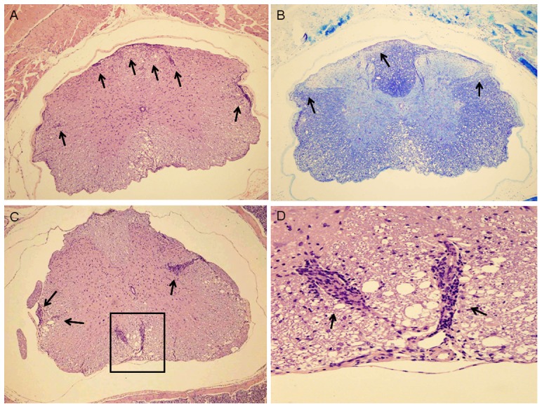
Figure 5. Histological analysis of cervical spinal cord (mouse #1-1).

A. Microscopic changes in transverse section of spinal cord are shown (100×). Multiple inflammatory foci and some multifocal inflammatory lesions are present in the leptomeninges, around blood vessels in the leptomeninges and white matter, and parenchyma of the white matter (arrows). There is also vacuolation in the white matter that is consistent with edema and demyelination. B. Luxol fast blue stained section of cervical spinal cord (100×). This section is from the same block of tissue as the H&E stained section shown in A. Areas of demyelination are visible within white matter (lighter blue stained areas, in the outer parts of the spinal cord; arrows). C. H&E stained transverse section of thoracic spinal cord (100×). Four multifocal inflammatory lesions are present in the leptomeninges, around blood vessels in the leptomeninges and white matter (box and arrows). There also is vacuolation in the white matter that is consistent with edema and demyelination (box). D. H&E stained section of thoracic spinal cord (400×). Detail of the above slide (boxed area). Two multifocal inflammatory lesions are shown (arrows).
