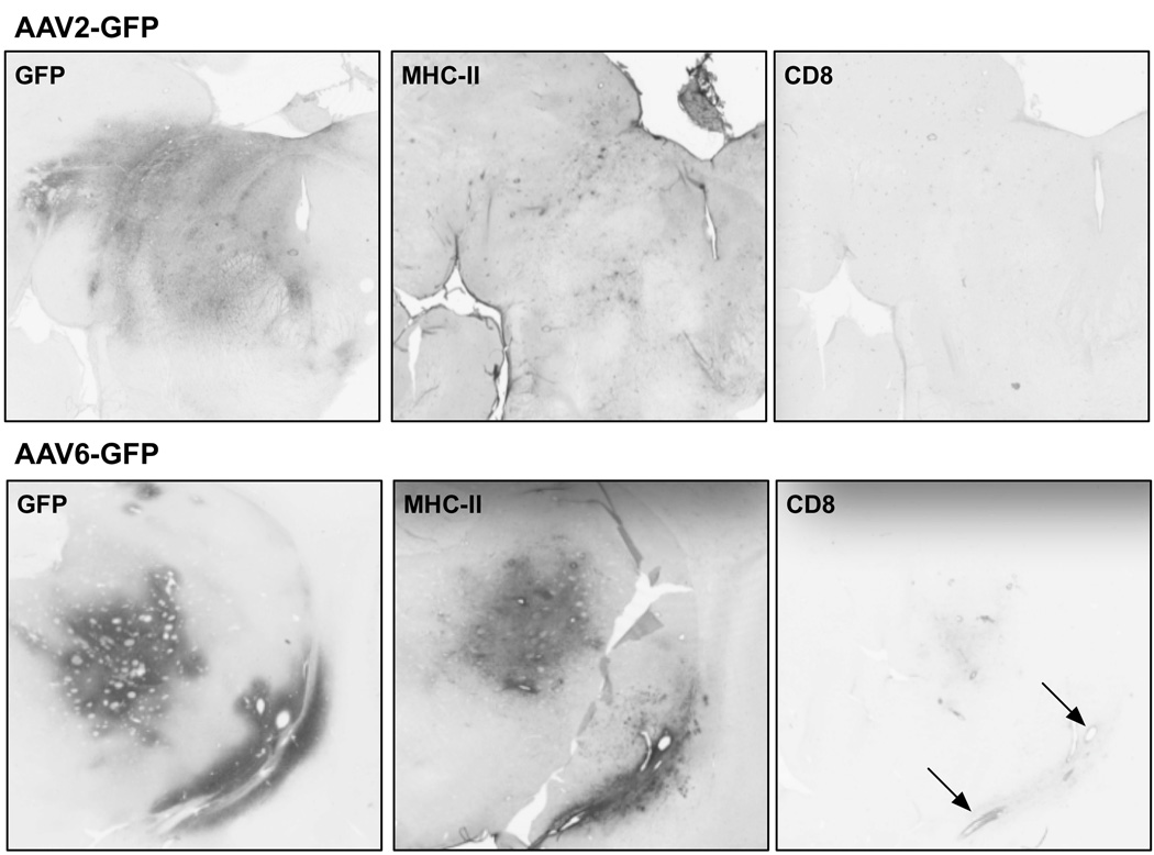
Figure 4. Induction of MHC-II expression by AAV6-GFP.
Scans of stained coronal sections of NHP brain at sites of infusion of AAV2-GFP (thalamus) or AAV6-GFP (putamen) stained for GFP, MHC-II and CD8. AAV2, because it transduces only neurons, does not elicit an immune response indicated by lack of MHC-II upregulation and absence of CD8-positive lymphocytes. In contrast, AAV6-GFP does trigger induction of MHC-II near the infusion site, presumably because it transduces non-neuronal cells in NHP. Also, some slight infiltration of CD8-positive lymphocytes is evident around blood vessels indicated by the arrows.

