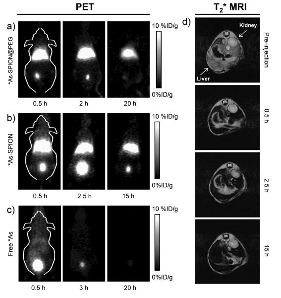
Figure 3.
Serial in vivo PET images of (a) PEGylated *As-SPION, (b) non-PEGylated *As-SPION and (c) free *As at different time points after intravenous injection into mice. (d) In vivo T2*-weighted MR images of mice before and after intravenous injection of SPION@PAA (in PBS). Transaxial images were presented to show the liver uptake of SPION@PAA.

