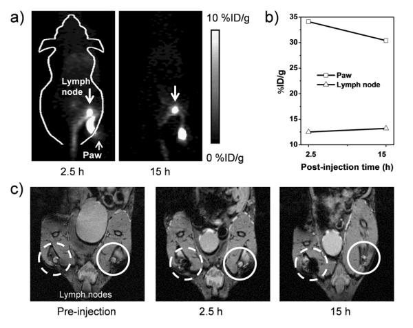
Figure 4.
(a) In vivo lymph node imaging with PET, after subcutaneous injection of *As-SPION@PEG into the right footpad of mouse. (b) Quantification of *As-SPION@PEG uptake in the lymph node and mouse paw. (c) In vivo lymph node mapping with MRI before and after injection of SPION@PAA to the left footpad of mouse. Obvious darkening of the lymph node could be seen (dashed circle) where the contralateral lymph node (solid circle) showed no contrast enhancement.

