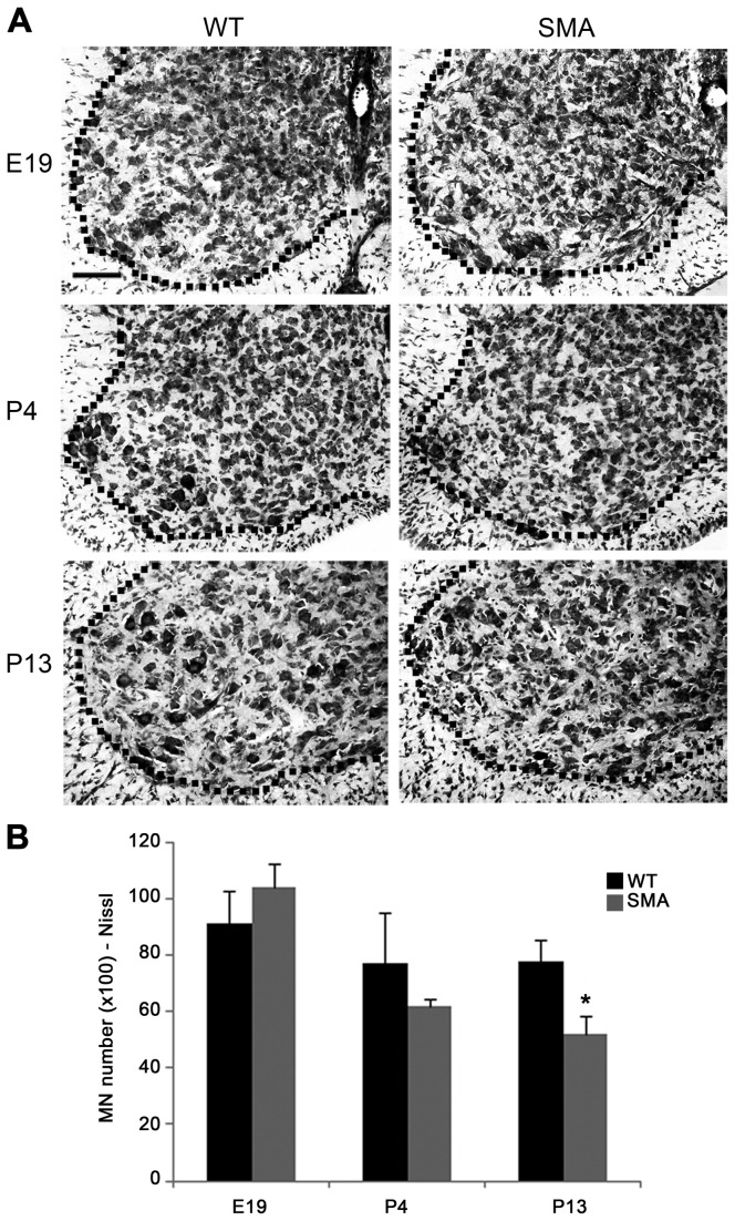
Figure 2. Stereological analysis of Nissl-stained motor neurons.
(A) Representative Nissl-stained coronal sections of ventral C5 spinal cord from WT (left) and SMA (right) mice at prenatal (E19, upper), pre- (P4, middle) and late-symptomatic (P13, lower) disease stages. (B) No difference in the total cervical motor neuron number was evident at prenatal stages (E19, left), whereas a progressive reduction was evident post-natally (P4, middle), reaching statistical significance at late-symptomatic stages (P13, right: *p<0.05, t-test). Values are expressed as mean ± SEM. Scale bar: 100 µm.

