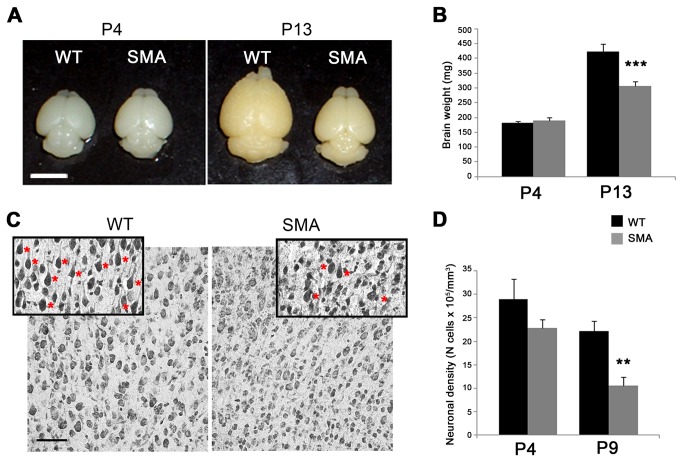
Figure 6. Loss of layer V pyramidal neurons of motor cortex.
(A) Photographs of P4 and P13 mouse brains showing a clear reduction of brain size in SMA vs WT mice at P13 (right). No difference in size were observed at P4 (left). (B) Quantitative analysis of brain weight in SMA vs WT littermates at P4 and P13. A weight reduction of about 30% was observed at late-symptomatic (P13: ***p<0.001, t-test) but not at pre-symptomatic (P4) stages. (C) Nissl-stained brain sections illustrating reduced numbers of larger pyramidal neurons (red asterisks in the inset) in layer V of SMA mice vs WT controls. (D) Stereological counts of Nissl-stained brain sections revealing a significant 52% decrease (**p<0.01, t-test) of pyramidal neuron density in layer V of P9 SMA vs WT mice. The density of pyramidal neurons was already reduced at P4, without reaching statistical significance (p>0.05). Values are expressed as mean ± SEM. Scale bars: 5 mm in A; 50 µm in C.

