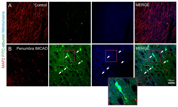
Figure 6. MCAO followed by reperfusion leads to the generation of ROS in cerebral microvessels.
Images from a representative control (A) hemisphere and its contralateral tMCAO hemisphere (B) of the neocortex are shown. MAP 2 (red), Fluorescein isothiocyanate –albumin (FITC-albumin; green) and anti-nitrotyrosine (blue) are displayed (n = 3 brains). Perfused control hemispheres (contralateral to tMCAO) displayed an intact MAP2 immunoreactivity, no FITC-albumin extravasation and absence of nitrotyrosine formation (A). In the penumbra of corresponding tMCAO hemispheres MAP2 damage was present as well as large and diffuse FITC-albumin extravasation. Here we found that cerebral microvessels (indicated by arrows and inset) were immunoreactive for nitrotyrosine indicating the formation of ROS (B). Note the cytosolic staining pattern of nitrotyrosine indicating the cytosolic localization of ROS.

