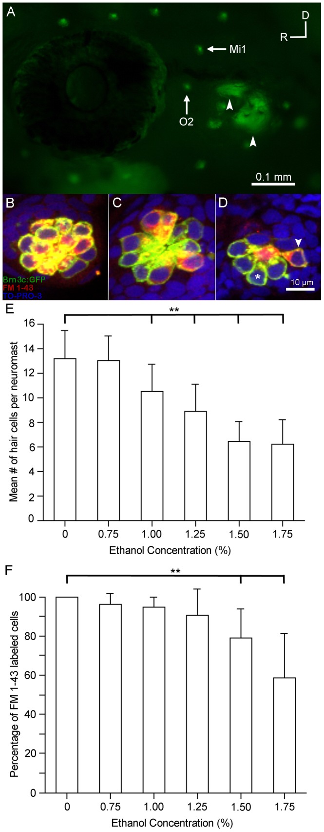
Figure 2. Ethanol but not methanol treatment affects the number of functional sensory hair cells in the lateral line.

Brn3c-GFP transgenic zebrafish embryos were raised in embryo medium supplemented with varying concentrations of ethanol. Prior to fixation, the larvae were stained with FM 1-43FX (red) to determine if hair cell mechanotransduction channels were functional. (A) Lateral view of a Brn3c-GFP zebrafish. Clusters of bright green GFP-labeled hair cells are found in neuromasts along the head and body of the animal. The O2 and Mi1 neuromasts are highlighted along with inner ear organs (small arrows). Higher magnification of hair cells of the O2 neuromast taken from z-stacks show that (B) hair cells in untreated controls and (C) 1% ethanol-treated animals had no observable morphological differences though there were fewer hair cells in the 1% ethanol-treated animals. Almost all of the GFP-labeled hair cells were co-stained with FM 1-43FX. (D) Fewer sensory hair cells were observed in neuromasts of animals treated with 1.5% ethanol. There were also fewer double-labeled cells (arrowhead) and more single labeled cells (asterisk). (E) Stereocilia bundles were counted to determine the number of hair cells present in the O2 neuromast. Control numbers were similar to those reported in other studies [23], [31]. Significantly fewer hair cells (**p<0.01) were counted in ethanol-treated larvae at concentrations greater than 1% per volume when compared to controls. No differences in the number of hair cells were observed in larvae treated with 1.5% methanol. (F) The percentage of GFP-labeled cells that were also co-stained for FM 1-43 decreased as the concentration of ethanol increased but not in methanol-treated larvae. There was a significant decrease (**p<0.01) in the number of double-labeled hair cells at the two highest concentrations of ethanol tested. Results are the mean values ± SD. n = 8-21 per condition.
