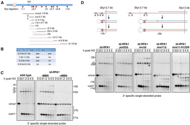
Figure 5. RPA prevents the formation of fold-back structures.
A. Schematic showing alkaline gel analysis of genomic DNA digested by StyI. Possible DNA intermediates formed after HO cutting and resection are shown. The red bars show the locations of the probes used. The red lines represent fragments detected by the 3’ strand specific probe close to the HO cut site. B. Four different dsDNA probes were used to determine the range of fragments r1b and r2b. C. Alkaline electrophoresis of the StyI-digested genomic DNA from different time points after HO induction. ssDNA intermediates were detected by the 3’ strand specific RNA probe. r1b and r2b bands can be eliminated by mung bean nuclease treatment of StyI-digested DNA. D. Schematic showing the formation of hairpins. Alkaline electrophoresis of the StyI-digested genomic DNA from different mutants showing their contribution to hairpin formation. See also Figures S3 and S5

