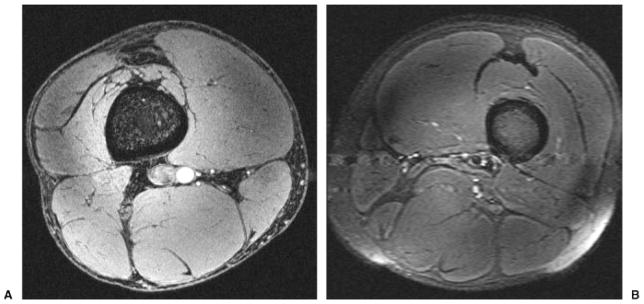
Figure 1.

(A) Axial T1-weighted 7-T magnetic resonance (MR) image of the thigh from a healthy male volunteer (3D gradient-recalled echo; TR/TE, 20 ms/4 ms; field of view [FOV], 13 cm × 13 cm; matrix, 512 × 512, 0.253 mm × 0.253 mm; slice thickness, 1 mm). Incidental note is made of flow-related enhancement within the popliteal artery. (B) Axial T2-weighted 7-T MR image of the thigh from a healthy male volunteer (turbo spin-echo; TR/TE, 3000 ms/60 ms; FOV, 16 cm × 16 cm; matrix, 256 × 256, 0.625 mm × 0.625 mm; slice thickness, 2 mm) in a different subject. There is pulsation artifact and coil inhomogeneity artifact near the periphery of the FOV.
