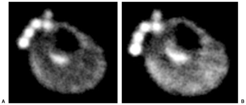
Figure 6.

(A) Resting axial 23Na 7-T magnetic resonance (MR) image from a healthy male volunteer demonstrates uniform signal intensity in the muscles of the calf. At the anterior aspect of the leg, phantoms containing NaCl of different concentrations (100 mmol, 150 mmol, 200 mmol, 250 mmol, and 300 mmol) are seen. (B) After 3 minutes of plantar flexion exercise, 23Na 7-T MR image demonstrates increased signal intensity within the posterior compartment muscles of the calf (gastrocnemius and soleus) used in the exercise. At the anterior aspect of the leg, phantoms containing NaCl of different concentrations (100 mmol, 150 mmol, 200 mmol, 250 mmol, and 300 mmol) are seen.
