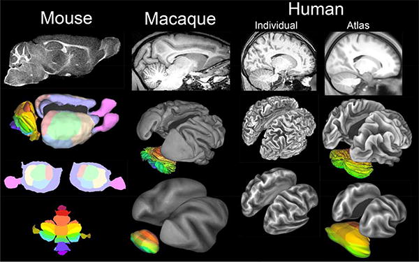
Figure 1.

Volume and surface representations of mouse, macaque, and human brains. Top row: Parasagittal slices of high-resolution T1w scans from 3 species. The mouse and macaque data are described in Van Essen (2002a,b). The human individual and group average (120 subjects) are from HCP scans acquired at high spatial resolution (0.7 mm) rather than the 1 mm isotropic voxels conventionally used, Lower panels: Surface reconstructions are shown as midthickness surfaces (all 3) and inflated surfaces (flatmaps for the mouse). Cortical lobes are colored on the mouse surfaces; cerebellar lobules are colored for all 3 species. The mouse surface includes the olfactory bulb. The HCP surface reconstructions benefitted from refinements to standard FreeSurfer processing (Glasser et al., 2013b; Glasser et al., 2013c). The human cerebellar surface is from the Colin individual MRI atlas (Van Essen, 2002b).
