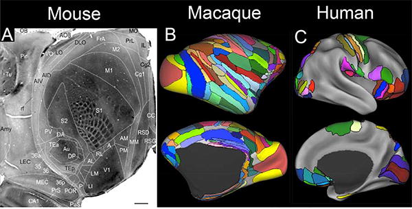
Figure 2.

Parcellations of mouse, macaque, and human cortex. A. A 40-area parcellation of mouse cortex illustrated on a cytochrome-oxidase stained tangential section of flattened mouse cortex. (Reproduced, with permission, from Wang et al., 2012). B. A composite parcellation of macaque cortex (Van Essen et al., 2012a) showing 130 areas of neocortex and transitional cortex, based on architectonic schemes of Lewis and Van Essen (2000b), Ferry et al. (2000), and Paxinos et al., (2000) and displayed on the inflated F99 atlas surface. C. A composite parcellation of 52 cortical areas spanning ~1/3 of human neocortex, based on published architectonic and retinotopic maps and displayed on the inflated Conte69 atlas surface (Van Essen et al., 2012a).(Ferry et al., 2000; Paxinos et al., 2000; Lewis and Van Essen, 2000b)
