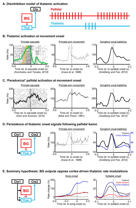
Figure 2. ‘Paradoxical’ co-activation of thalamic neurons and their inhibitory BG inputs, observed across model systems and behavioral paradigms.
(A) Left, the place of the thalamus in a canonical BG-thalamocortical loop. In a predominant view of BG-thalamocortical transmission, thalamic activation occurs during pallidal pauses, through a process of disinhibition. Right, schematic of pallidal and thalamic spike trains (red and blue, respectively), showing thalamic activation during a pause in tonic pallidal firing. (B–C) Movement-onset aligned rate histograms showing thalamic (B) or pallidal (C) activation at movement onset in primates during saccades and arm movements, as well at syllable onset in songbirds during vocal babbling. (D) Left, the BG-recipient thalamus is embedded in a cortico-thalamocortical loop. Movement onset-aligned rate histograms show that thalamic onset signals persist following lesion of pallidal inputs in primates (center) and in songbirds (right). In songbirds, antidromically identified corticothalamic neurons also exhibit rate peaks at syllable onsets. Together, these findings suggest that cortical inputs to the BG-recipient thalamus can drive thalamic pre-motor signals, and further suggest that BG inputs may oppose the cortically driven rate modulation. (E) Left, a schematic of the BG-thalamocortical loop highlighting that cortical regions projecting to the BG can also project to the thalamus [77, 78]. Rate histograms showing that pallidal (red), thalamic (black) and corticothalamic (blue) neurons exhibit rate increases prior to song onsets (center), as well as syllable onsets (right)(Data from Goldberg and Fee, 2012). Such coactivations might be explained by cortical inputs that project to both the hyperdirect pathway and to the thalamus. Panels in (C–E) are reproduced with permission from (Shin and Sommer, 2010; Mink and Thach, 1991; Goldberg and Fee, 2012; and Inase et al, 1996).

