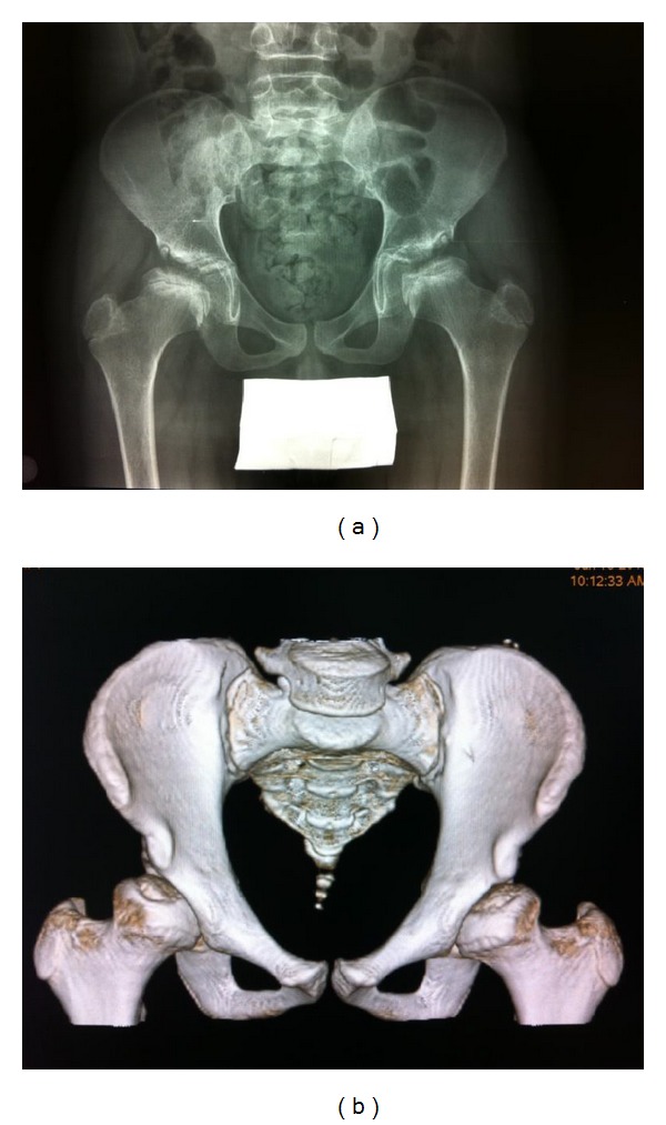
Figure 1.

(a) First pelvic X-ray: dysplastic appearance of both femoral heads and irregular upper contour of the acetabular roof with mild coxa vara. (b) Last pelvic CT scan (three-dimensional volume-rendered reformatted picture): almost total reconstruction of anatomic structures.
