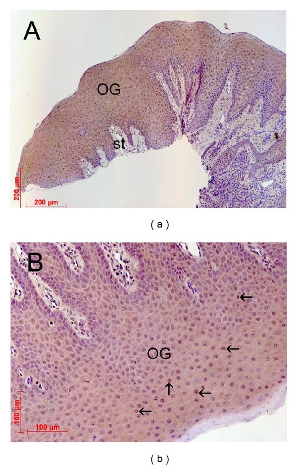
Figure 1.

Demonstration of the glycine receptor (GlyR) in gingival tissue. Overview (a) of oral tissue (magnification ×10) isolated from extracted upper third molars showing different region of the gingival tissue (OG: oral gingival tissue; st: subepithelial tissue). (b) Representative gingival tissue section (magnification ×200) assessed by immunohistochemistry using an anti-glycine receptor antibody (SYSY, Göttingen, Germany) at 4°C overnight and counterstaining with DAB (brown color). There was strong immune reaction in the epithelial layers of the gingiva, namely, in gingival keratinocytes. The underlying basal membrane as well as the connective tissue showed negative immune staining.
