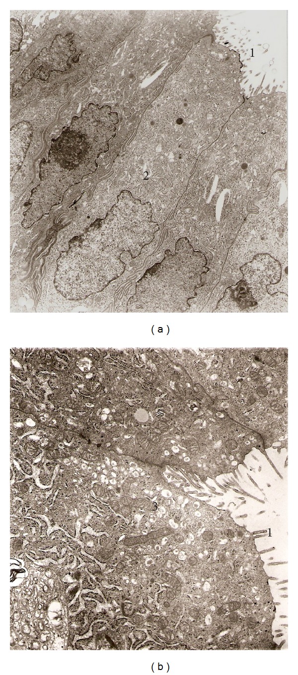
Figure 5.

Electron micrographs from rat uterine horn (Control). (a) Epithelial cells showing microvilli facing to lumen (1) and normal nuclear membrane (2), 4,980x. (b) In addition numerous secretory granules and mitochondria can be observed also (3), 8,800x.
