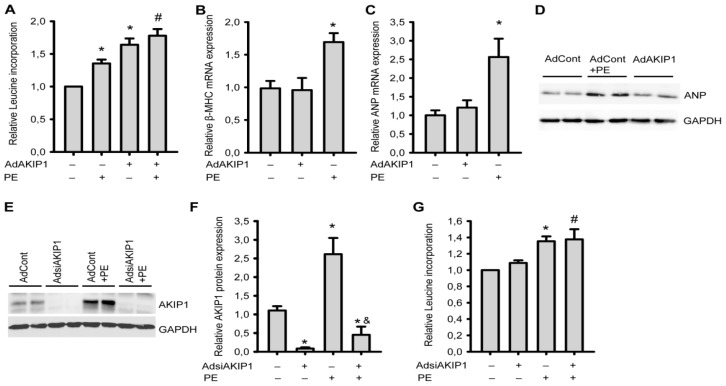
Figure 3.
AKIP1 and neurohormonal-induced hypertrophy. One day after isolation, NRVCs were infected overnight with the indicated adenovirus in the presence of serum and subsequently serum-starved for 24 h. The cells were then stimulated with PE (50 μM) for 24 h. (A) AKIP1 could further increase PE-induced hypertrophy (*p < 0.01 to AdControl group, n = 8; # p < 0.01 to PE group, n = 8); (B) and (C) AKIP1 does not trigger the re-expression of the fetal gene program. β-MHC (B) and ANP (C) expression in control, AKIP1 overexpressing and PE treated cells are shown. mRNA expression was normalized to Cyclophilin A (*p < 0.01 to control, n = 6); (D) ANP protein expression in control, AKIP1 overexpressing and PE treated cells, which is consistent with mRNA expression (representative blot is shown, n = 3); (E) A representative Western blot is shown of cells treated with siAKIP1 adenovirus in the presence or absence of PE and (F) Quantification of all western blots (*p < 0.05 to AdControl group; & p < 0.05 to PE group, n = 3); (G) siAKIP1 could not inhibit PE-induced hypertrophy measured by protein synthesis (*p < 0.01 to AdControl group, n = 8; # p < 0.01 to AdsiAKIP1 group, n = 8).

