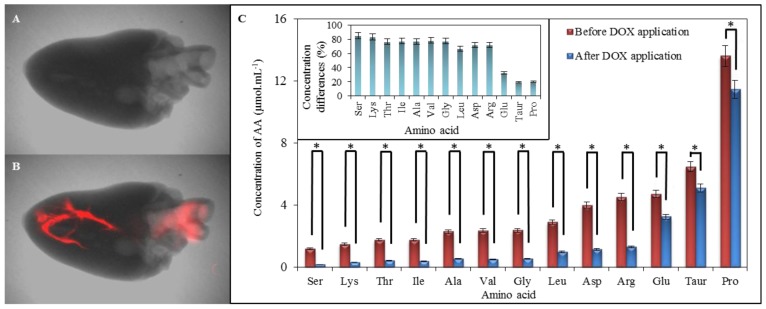
Figure 8.
Comparison of chicken myocardium before and after application of 1000 μg mL−1 of doxorubicin dissolved in physiological saline solution. (A) Chicken cardiac muscle tissue without doxorubicin applied (X-ray image with overlaid fluorescence image); (B) Chicken cardiac muscle tissue with 50 μL of doxorubicin applied (X-ray image with overlaid fluorescence image). The fluorescence of doxorubicin was detected by Carestream In Vivo Xtreme Imaging System; (C) Expression of IELC results of myocardium amino acids content analysis. Both, control and heart, after application of doxorubicin were obtained as the averages from ten measurements. In inset it can be seen the percentage expression of differences between AA concentrations of amino acids in myocardium between and after application of DOX. * refer the differences between amino acid contents as statistically significant (at the p = 0.05 level).

