Figure 43.

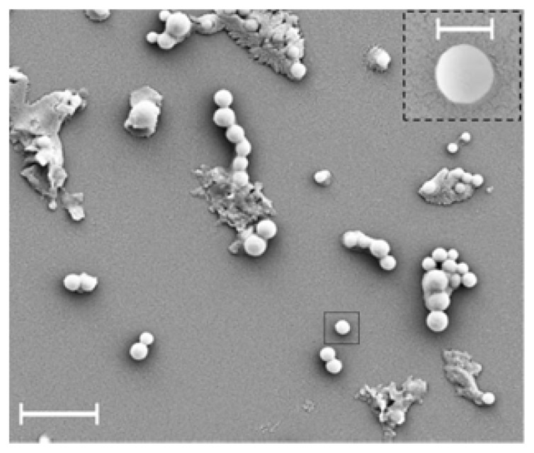
Scanning electron microscopy image of proteo-SLNs spread on a glass substrate (scale bars 1 μm and 200 nm (inset)) [120].

Scanning electron microscopy image of proteo-SLNs spread on a glass substrate (scale bars 1 μm and 200 nm (inset)) [120].