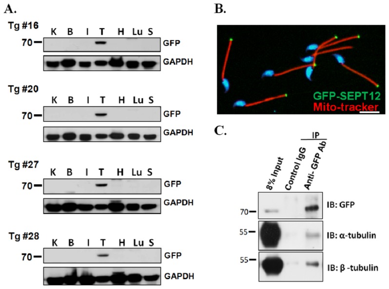
Figure 2.
Expression of SEPT12, α- and β-tubulin in the SEPTIN12-transgenic mice. (A) SEPT12 expression in different organs from the SEPTIN12-transgenic mice. The tissue lysate from kidney (K), brain (B), intestine (I), testis (T), heart (H), lung (L) and spleen (S) were analyzed by Western blotting with anti-GFP or anti-GAPDH antibody (loading control); (B) Spermatozoa isolated from vas deferens of SEPTIN12-transgenic mice were stained with mito-tracker (Red), DAPI (Blue) and SEPT12 (Green); Scale bar: 5 μm; (C) Co-IP of GFP-SEPT12 and α- or β-tubulin. Testicular lysates from SEPTIN12-transgenic mice were immunoprecipitated (IP) with anti-GFP antibody (lane3) followed by immuno-blotting (IB) with anti-GFP, anti-α-tubulin and anti-β-tubulin antibody, respectively. IgG was used as control. Input protein (8%) was used as control of immunoblotting (IB) in the testicular lysates.

