Figure 4.
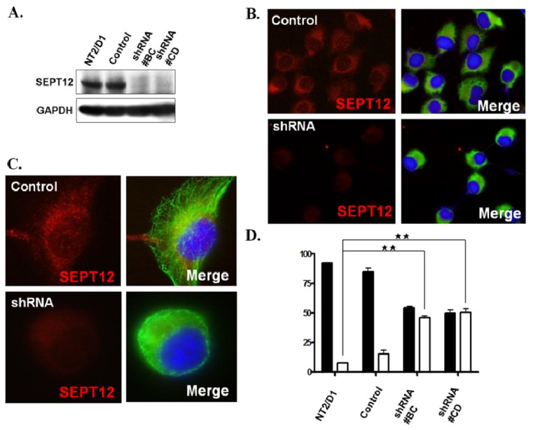
Silencing SEPTIN12 affects α-/β-tubulins organization in the NTERA-2 d.D1 cell. (A) Expression levels of SEPT12 in the stable clone derived from the NTERA-2 d.D1 cell (shRNA #BC and #CD for SEPTIN12). Control: a shRNA for the GFP sequence. Each lane was probed with the antibody against SEPT12 or GAPDH (as a loading control) in immuno-blotting; (B,C) Immuno-fluorescent detection of SEPT12 (Red) and α-tubulin (Green) in NTERA-2 d.D1 cells treated with shRNA for GFP (control) or SEPTIN12. Cells were stained with anti-SEPT12 (red, left panel) and merge of images for anti-SEPT12 and anti-α-tubulin (green, left panel). Magnification ×400 (B) and ×1000 (C); (D) Quantification of disorganized α-tubulin in the SEPTIN12 knockdown cell. There are two distinctive patters of α-tubulin in the SEPTIN12 knockdown cell: the polymerized form (black bar) and the disorganized form (white bar). The percentages of two distinct patterns were calculated by randomly selected cells (n = 300, duplication). Two-tailed Student’s t-test; Error bars indicate ± SEM (**, p< 0.001).

