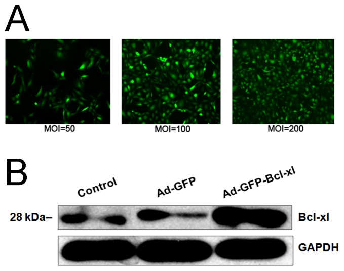
Figure 1.
HUVECs infection with recombinant adenovirus (Adv-GFP-Bcl-xl) or empty virus (Adv-GFP) and the expression of Bcl-xl by Western blotting. (A) Observation of HUVECs infected at different multiplicities of infection (MOI) for 48 h by fluorescence microscopy (×40). Green fluorescence represents infected cells; (B) Bcl-xl expression in HUVECs of different groups by Western blotting.

