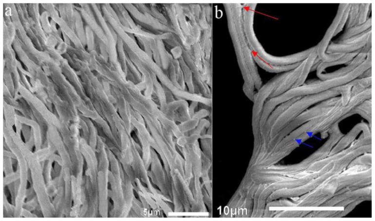
Figure 3.
Scanning electron microscopy (SEM) examination of artificial sclerotia at 8 °C and mycelia at 25 °C after 120 days of cultivation. (a) depicts loosened mycelia cultured at 25 °C without sclerotial development; (b) depicts the condensed and fused mycelia in the SM stage of sclerotial formation cultivated at 8 °C. Images are representatives of three independent experiments. Scale bar, (a) 5 μm; (b) 10 μm.

