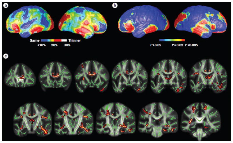
Figure 4.

Reduced gray matter thickness and white matter integrity in left MTLE. a | Mean percent reduction in cortical thickness as a percentage of control average. Red areas in the bilateral in the frontal poles, frontal operculum, orbitalfrontal, lateral temporal and occipital regions, and the right angular gyrus and primary sensorimotor cortex surroundings the central sulcus denote ≤30% decrease in thickness, on average, compared with corresponding areas in controls. b | Significance of these changes shown as a map of P values. c | Reductions in white matter integrity, measured by decreased fractional anisotropy, were evident in mesial and lateral temporal lobe, limbic system and extratemporal lobe regions, particularly ipsilateral to the side of seizure onset. Yellow and dark red regions indicate white matter tracts with decreased fractional anisotropy. Green regions indicate areas not notably different from controls. Only left MTLE patients are presented here, although similar gray and white matter abnormalities—albeit to a lesser degree—were evident in right MTLE. Parts a and b are modified, with permission from Oxford University Press © Lin, J. J. et al. Cereb. Cortex 17, 2007–2018 (2007). Part c is modified with permission from Elsevier Ltd © Focke, N. K. et al. Neuroimage 40, 728–737 (2008). Abbreviation: MTLE, mesial temporal lobe epilepsy.
