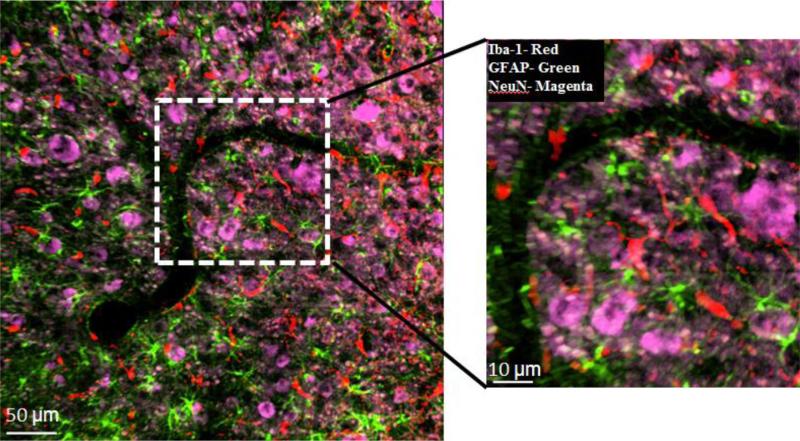
Fig. 5.
Quality of triple-label immunostaining after 24-h-treatment on the same 40 μm-thick cat brain specimen as shown in Fig. 4D, using primary antibodies against Iba-1 (microglia), GFAP (astrocytes), NeuN (neural cell bodies) and corresponding AlexaFluor® fluorophores: Iba-1 (488 nm), GFAP (633 nm), and NeuN (546 nm). Image was obtained on LSM 510 laser scanning system. False colors were applied for each fluorophore. Iba-1, red; GFAP, green; NeuN, magenta. (For interpretation of the references to color in this figure legend, the reader is referred to the web version of this article.)

