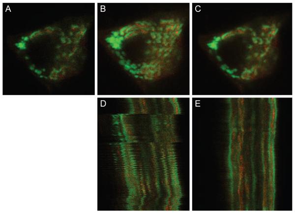
Figure 5.
(A) Example image from salivary gland data set. (B) Maximum projection of 15 images before registration. (C) Maximum projection of 15 images after registration. (D) Line scan projection image before registration. (E) Line scan projection image after registration. Image field before registration is 25 microns wide. Image field after registration is wider because the field has shifted over the time of collection to include more area. Image sequence contains 310 frames. Non-rigid registration was performed using λ = 0.005 and a B-spline control point grid spacing of δx = δy = 16 pixels, and using a limited memory Broyden-Fletcher-Goldfarb-Shanno (LBFGS) optimizer to determine the final B-spline control points.

