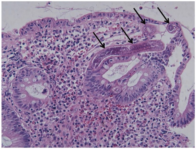
Fig. 3.

Photomicrograph of the biopsy specimen from the distal ileum (H&E stain, ×200). Biopsy specimens of the ileum show atrophic changes of the intestinal villi, infiltration of plasma cells and eosinophils, and several sections of worms invading the mucosa (arrows).
