
Figure 2.
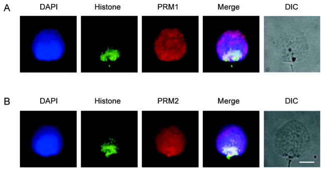
Immunofluorescent colocalization of the core histones with protamine 1 (PRM1) and protamine 2 (PRM2) in permeablized and decondensed human sperm cells. (A): Immunofluorescent colocalization of histones with anti-core histones antibody plus PRM1 with anti-protamine 1. (B): Immunofluorescent colocalization of histones with anti-core histones antibody plus PRM2 with anti-protamine 2. The corresponding grey-scale DIC image of the intact permeablized cells is shown. DNA stained with blue 4′-6-diamidino-2-phenylindole (DAPI), histones green, with Alexa Fluor 488-conjugated streptavidin and protamines red with CY5-conjugated goat anti-mouse antibody. The merged pseudo-color image is shown in the right most panel. Scale bar=5 μm. Note that core the histones localize towards the post acrosomal posterior ring region end while PRM1 and PRM2 occupy the entire nucleus.