
Figure 1. Representative Immunohistochemical staining of IL-17A expression in normal and breast cancer human tissues.
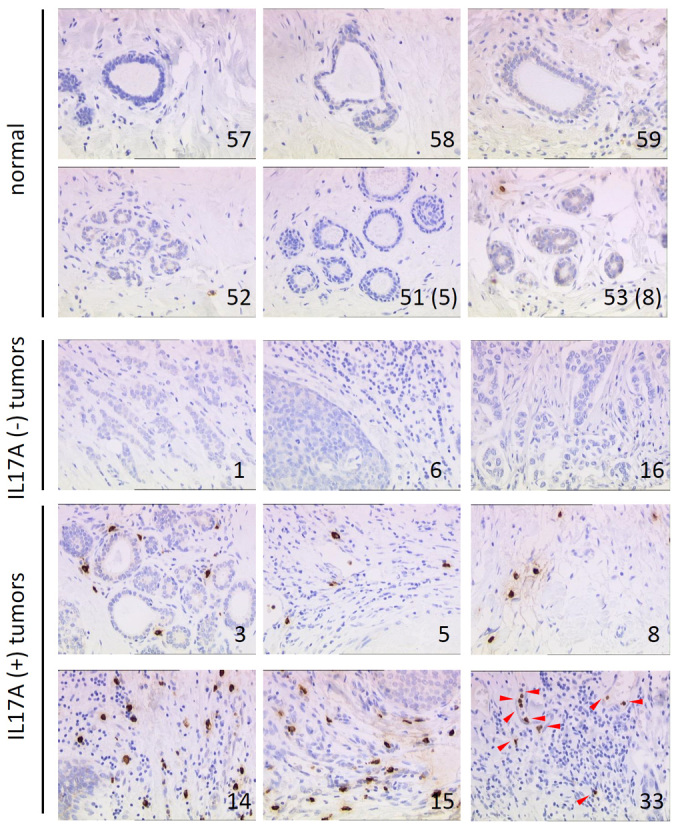
IL-17A stained sections of 40 invasive ductal breast carcinomas, 10 metastases and 10 matched normal counterparts. Brown staining indicates IL-17A protein. Arrows indicate IL-17A positive cells within the stroma, # refers to the corresponding sample in Supplementary Table 1.