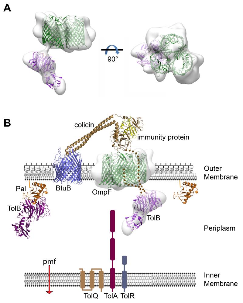
Figure 4. ColE9 uses OmpF to capture TolB on the other side of the membrane in a defined orientation.
(A) Negative stain EM structure at ~20 Å resolution of the trypsin digested ColE9 translocon comprising OmpF trimer and TolB connected by ColE9 T2-122, seen in side (left) and face views (right) of the membrane plane. OmpF and TolB crystal structures have been manually docked into the density as rigid bodies. (B) Structural representation of the ColE9 translocon – the structure for the related ColE3-Im3 bound to BtuB is shown (pdb accession code 1UJW) – and its protein-protein interaction network across the Gram-negative cell envelope (see text for details) (14, 21)

