Figure 1.

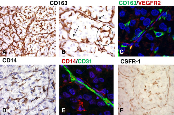
Macrophages are key components of the inflammatory microenvironment in metastatic alveolar soft part sarcoma. Immunohistochemical analysis of CD163+ cells infiltrating an untreated ASPS lesion (A-B). As evidenced by the higher magnification image these cells are found in two distinct localizations: they are interspersed within nest tumor cells (circle) and they are also detectable in the perivascular region (arrows). (B) Confocal microscopy imaging of CD163+ cells (green) shows that they are aligned to endothelial VEGFR2+ cells (red) (C). CD14 staining closely resembles that of CD163 (D) and double staining confirms that CD31+ endothelial cells (green) are lined by CD14+ macrophages (red). (E) A similar distribution of immunoreactivity is observed for CSFR-1 (F).
