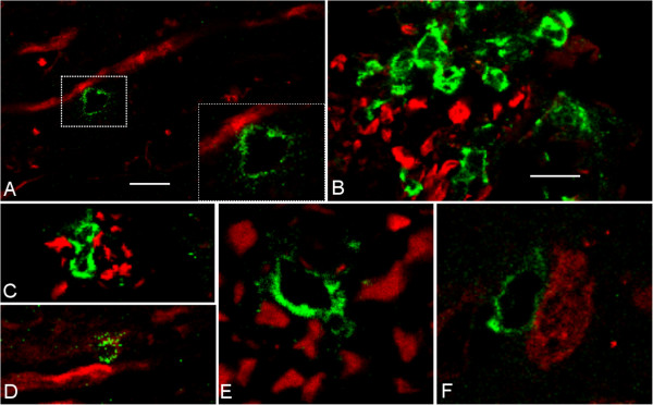
Figure 2.

T cells associated with axonal damage in spastic mice. Confocal images of (A) CD3+ T cell (green) in contact with SMI32+ axon (red). (B, C) CD4+ T cells in area of axonal damage and (D) next to swollen NF-L+ axons. (E) CD8+ T cells between NF-L+ axons and (F) Neu-N+ neurons in the spinal cord. A, E, F, bar = 5 μm; B, C, D, bar = 25 μm.
