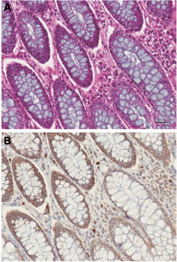
Figure 1.

Immunohistochemistry for PP6 in normal human colon. Sections were stained with hematoxylin and eosin (A) and immunostained with anti-PP6 using peroxidase to give brown color (B). The droplets of mucosal fluid in goblet cells appear blue in (A) and white in (B) and these structures are surrounded by the continuous layer of epithelial cells with high levels of PP6. The scale bar represents 10 um. Multiple sections from two independent specimens were stained and examined.
