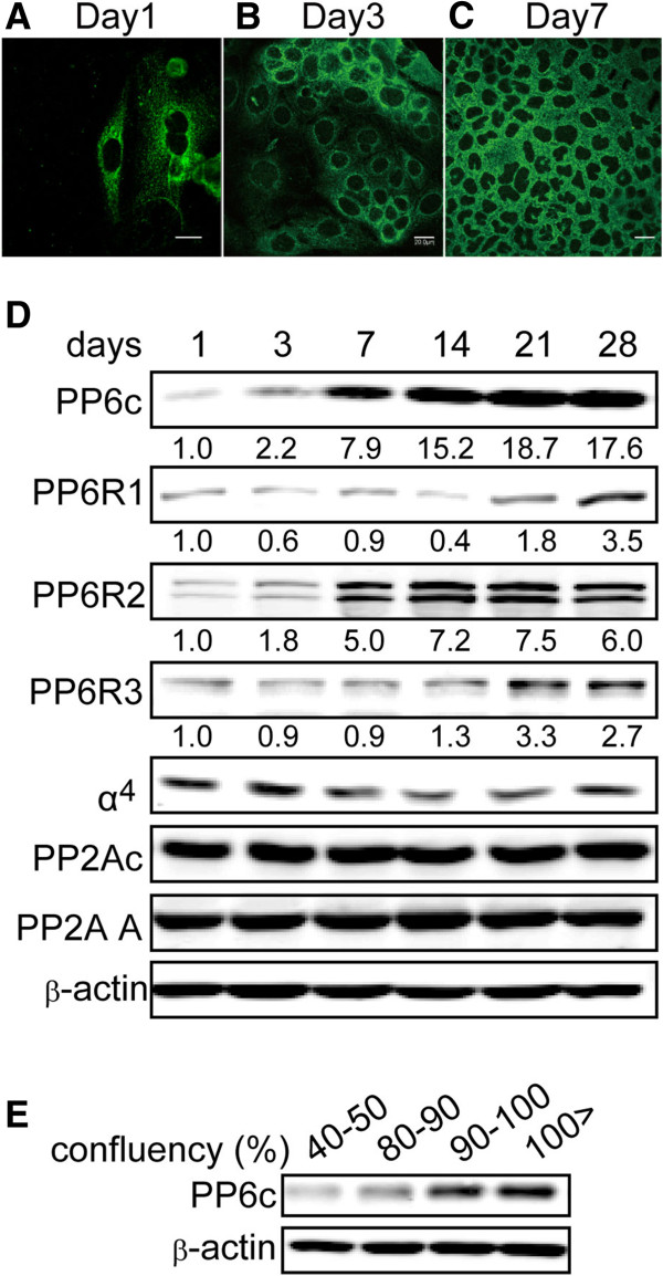
Figure 2.

Accumulation of PP6 protein in high density epithelial cells. (A-C) Caco-2 cells were cultured on Nunc Lab-Tek II Chamber slide 4 well (72,000 cells/ well) and cultured for 1 (A), 3 (B) and 7 (C) days. Cells were fixed, permeabilized and stained with anti-PP6 antibodies and images captured with an Olympus confocal microscope. Similar results were obtained in 3 independent experiments. (D) Caco-2 cells were cultured for indicated time periods and extracts analyzed for the indicated proteins by immunoblotting. Numbers below frame are the relative fluorescent intensity of PP6c staining determined with an Odyssey 2D scanner (Licor Industries). Images are representative of two independent experiments. (E) Different numbers of human ARPE19 epithelial cells were seeded and after 4 days confluency estimated from microscopic examination (as indicated). Levels of PP6 protein were determined by immunoblotting, with actin as a loading control for total protein. Images are representative of two independent experiments.
