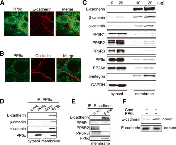
Figure 4.
Association of PP6 with Adherens Junction complexes. Caco-2 cells were immunostained for (A) PP6 (green) and E-cadherin (red), with yellow showing extensive overlap and (B) PP6 (green) and occludin (red) with essentially no co-localization. Confocal microscope images are selected from 3 independent experiments that had the same results. (C) Cytosolic and membrane fractions of Caco-2 cells were separated by sucrose gradient, and indicated proteins detected by immunoblotting samples of 10 or 20 μg total protein. Images are representative of two independent experiments. (D) Endogenous PP6c was immunoprecipitated from cytosolic and membrane fractions of Caco-2 cells in parallel with samples using non-immune rabbit IgG as control. Immuncomplexes were resolved by SDS-PAGE and immunoblotted for PP6c, E-cadherin, and α and β cateninin. Images are representative of two independent experiments. (E) E-cadherin was immunoprecipitated from the membrane fraction, and normal rabbit IgG used as control and samples immunoblotted for recovery of E-cadherin, PP6c, PP6R1, PP6R2 and PP6R3. Images are representative of two independent experiments. (F) S-tagged PP6c and empty vector control (Cont.) were used in pull-down assay with 35S radiolabeled cytosolic domain of E-cadherin expressed in a cell free system, as described. The bound and unbound proteins were resolved by SDS-PAGE and the gel used for exposure of a PhosphorImager plate to compare the amounts of bound E-cadherin domain. Images are representative of two independent experiments.

