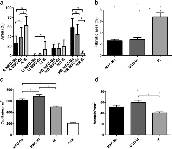
Figure 4.
Morphometric analyses of limb muscles. From Figure 3, (a) necrotic, regenerative and normal areas, (b) fibrotic area, (c) capillary density and (d) matured vessels were determined. More than 50 fields of lesions were counted for each group. A, adipocyte; Is, ischemic group; LI, Infiltrated leukocyte; MD, muscle degeneration; MR, muscle regeneration; MSC-Ba, ischemic animals treated with MSCs obtained from BALB/c mice; MSC-Bl, ischemic animals treated with mesenchymal stem cells (MSCs) obtained from C57/BL6 mice. *P <0.05.

