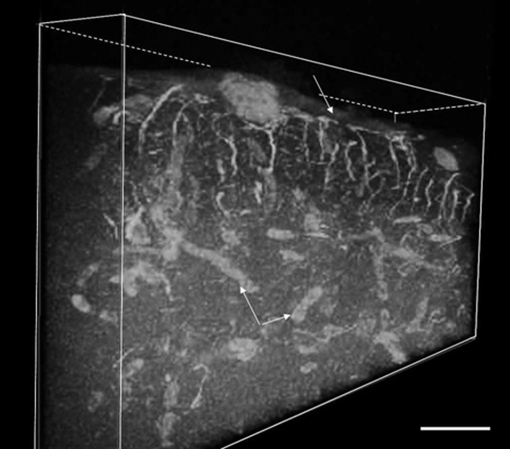
Figure 10.
Three dimensional representation of 30 µm thick stratum fibrosum marginale stained with GFAP immunohistochemistry and collected from an 80 µm thick vibratome section. Note the richly arranged, radially orientated GFAP positive astrocytic fibers with the layers of end feet attaching to the pia mater (white arrow), creating the glial limitans. Double arrows with the asterisk point toward the labeled blood vessels. Micron bar = 30 µm.

