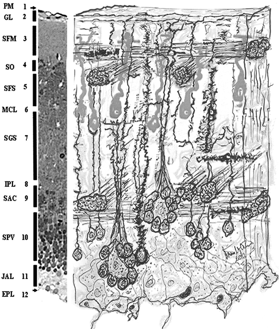
Figure 11.
Composite drawing of the key features seen in the optic tectal cortical structure. The photographic strip at the left of the figure and the naming of the layers is taken from Figure 2 as a guidance to the diagram. In an enhanced way, the rosettes are depicted as they are seen in the SPV (10). A different cell central to the neuronal cell bodies that form rosettes is also shown. The dendritic branches of these structures are grouping together forming structures like a bunch of scallions. This picture also enhances the juxtaventricular aqueous layer and the monolayer of the magnocellular neurons (6).

