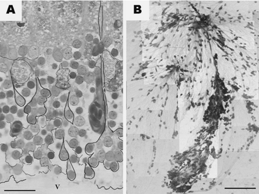
Figure 4.
Periventricular projecting neurons in a toluidine blue stained semithin section (A) and in a toluidine blue stained squashed preparation (B). In panel (A), black contours are labeling different periventricular projections neurons either as single cells or as cells grouped (bunched) into what we call rosettes. Also note the large, fluid-filled appearing spaces between the periventricular cells. V - tectal ventricle. (B). The dendrites of several neuronal bunches, of varying size, are joining in the upper part of the picture. Both images are montages of several independent photographs captured with the 100× oil objective. Micron bar: A = 150 µm; B = 30 µm

