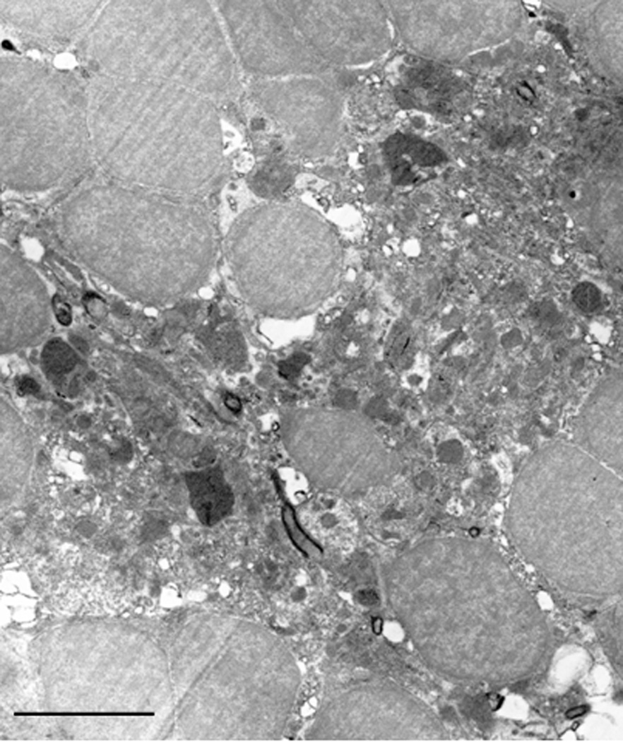
Figure 6.
Electron micrograph of the rosette-like arrangement of the periventricular projecting neurons. The electron-dense cell located in the center of the picture is surrounded by the periventricular neurons forming a basket. Note the even distribution of the hetero and euchromatin in all cell type nuclei and the very small ring-like cytoplasm of the neurons. The cytoplasm of the centrally located cell is full of organelles such as vesicles which could be lysozomes or perioxisomes as well as mitochondria. Micron bar = 10 µm

