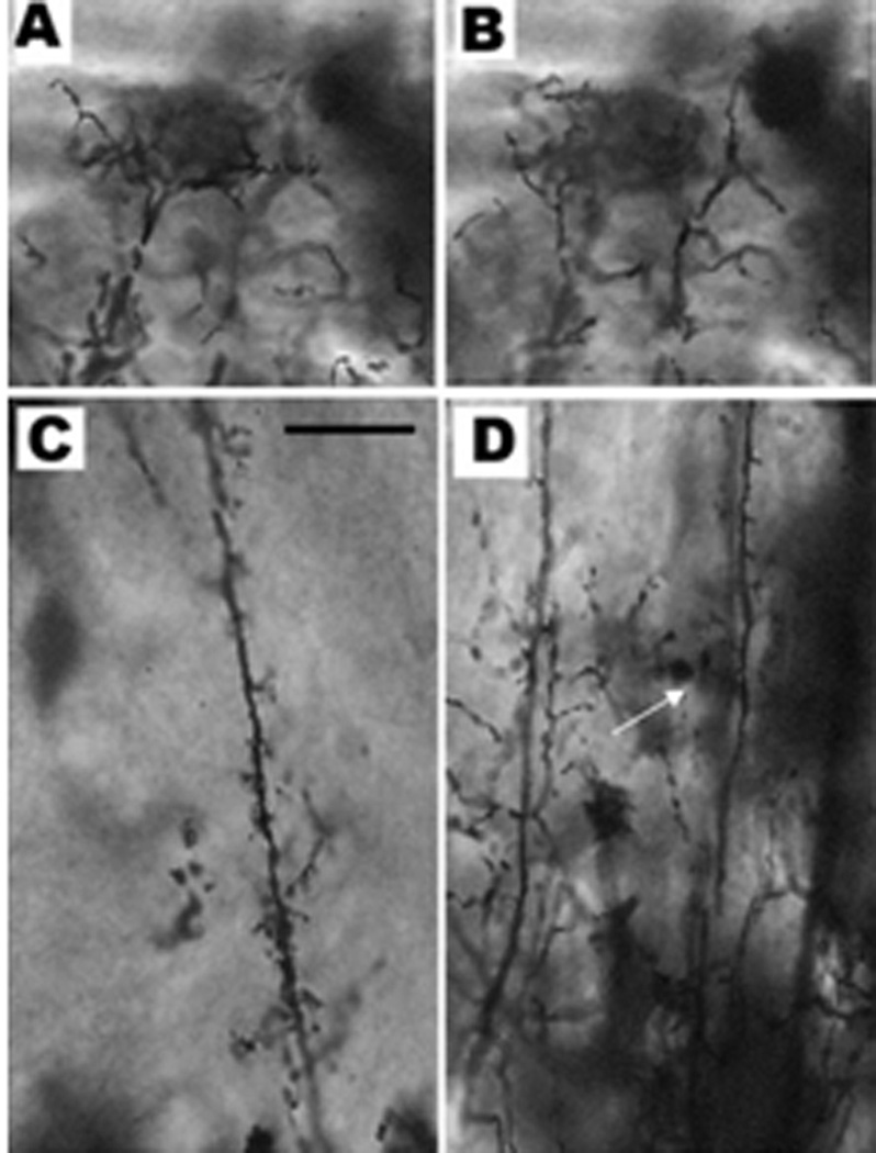
Figure 7.
Periventricular neurons stained with rapid Golgi. A & B depict dendritic arborization in the stratum fibrosum marginale of all types of neurons. C & D are different mid-regions of periventricular neuronal dendrites as seen in the stratum fibrosum & gresium superficale. C corresponds to the monostratified periventricular interneuron (the dendrites divide only at the marginal layer). D depicts two types of periventricular neurons: in the left of the picture is a typical periventricular projection neuron with numerous short side dendritic branches seen in many or all of the cortical layers. In the right of the picture is the bi-stratified projetion neuron which branches not only in the marginal region but also in the stratum fibrosum (white arrow). Micron bar = 20 µm

