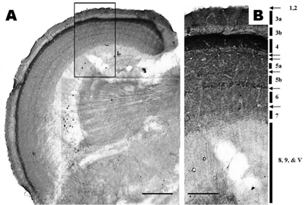
Figure 8.
A. Nestin immunohistochemical stain of neuronal components as seen in an 80 µm thick vibratome section of the adult zebrafish optic tectum. Scale Bar = 150 µm. B. A montage of the framed area in image A captured with 100× oil immersion objective.
1. Pia mater; 2. Glia limitans; 3a & b. Stratum fibrosum marginale; 4. Stratum opticum; 5a & b. Stratum fibrosum and grecium; 6. Stratum album central; 7. Stratum periventricular; 8 & 9. Stratum periventricular; V. Tectal ventricle. Note the intensively stained synaptic layers (arrows) in layers 4,5, & 6. Micron bar = 100 µm

