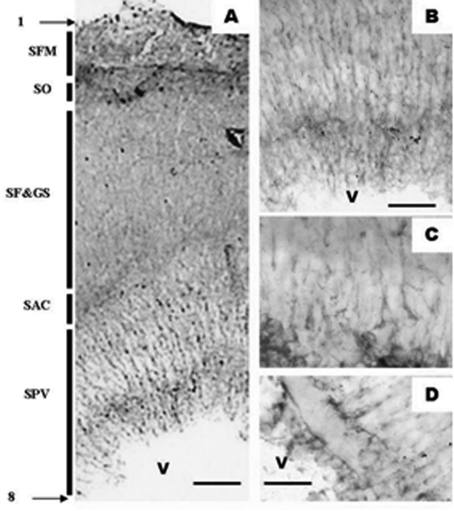
Figure 9.
Adult zebrafish optic tectum as seen in a anti-GFAP-DAB reaction in an 80 µm thick vibratome section. A. is a general overview as seen under a 10× objective B. The periventricular gray zone as seen under 40× objective. C & D. are the periventricular gray zone with 100× oil immersion. Note that the layering of the optic tectum seem in GFAP differs from what we have observed in toluidine blue and nestin staining. 1. Pia mater; SFM - Stratum fibrosum marginale; SO - Stratum opticum; SF&GS - Stratum fibrosum and griseum superficiale; SAC - Stratum album centrale; SPV - Stratum periventriculare; 8. Ependyma; V - Tectal ventricle Micron bars: A = 60 µm , B = 30 µm , C&D = 20 µm

