
Figure 2.
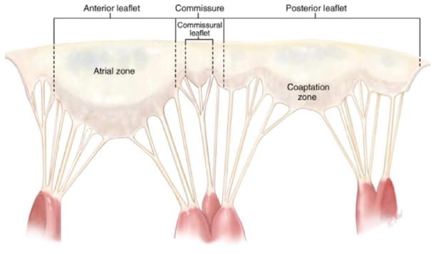
The mitral valve is unfolded and the atrial leaflet surface exposed. The papillary muscles have been dissected and the heads remain attached via chordae tendineae to the anterior, posterior and commissural leaflets (Adapted from Carpentier A et al. Carpentier’s Reconstructive Valve Surgery. From Valve Analysis to Valve Reconstruction. 2010 Saunders Elsevier)