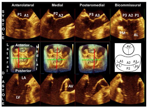
Figure 4.
The middle panel shows a 3D-rendered surgical view of the mitral valve. A schematic on the right side of the middle panel helps identify the mitral valve scallops and aortic valve in this view. The upper panel shows 2D Echo views of precisely known orientation because they are derived as slices of the 3D mitral valve apparatus in systole. The slice plane is indicated by the red line in the middle plane (perpendicular to a bicommissural axis, green line). The lower panel shows the same slice planes in diastole with the mitral leaflets open. (AL, anterolateral papillary muscle; AV, aortic valve; LA, left atrium; LV, left ventricle; PM, posteromedial papillary muscle)

