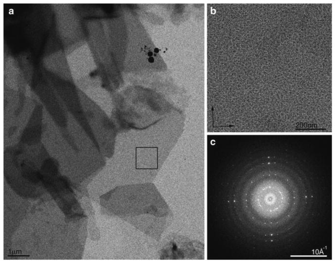
Fig. 3.

Cryo EM of two-dimensional crystals. (a) Crystals of the water channel aquaporin-0 are large and have sharp edges attesting to the degree of order within. (b) High-resolution image of the crystal area highlighted by a box in (a). (c) Fourier transform of the image in (b) showing strong and sharp spots to ∼6Å resolution. These crystals are ready for analysis by electron diffraction because the crystals appear uniformly grey on the grid. The spots in the Fourier transform are sharp and extend to ∼6Å resolution without unbending. At this stage the sample should be frozen and the microscope setup should be changed to diffraction and data collected.
