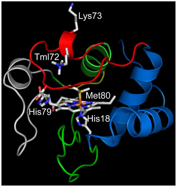
Figure 1.
Structure of yeast iso-1-Cytc showing the K79H mutation. The pdb file 2ycc was used and the K79H mutation inserted in silico (HyperChem). The side chains of Lys73 and trimethyllysine 72 (Tml72) and the heme and its native state ligands, His18 and Met80, are shown as stick models colored by element. The heme crevice loop (Ω-loop D, residues 70 to 85) is shown in red.

