
Fig. 2. Confocal immunofluorescence microscopy of epitope-tagged wild type and mutant δ opioid receptors expressed in CHO cells.
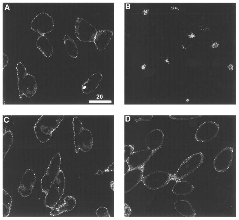
CHO cells expressing wild type (A and B) or ΔC15 receptor (C and D) were incubated in the absence (A and C) or presence of 100 nM DADLE (B and D) for 30 min. Fixation, permeabilization, and immunofluorescence staining of the receptors with the monoclonal antibody against the epitope tag are as described under “Experimental Procedures.” Cells were imaged by confocal fluorescence microscopy, using a plane of focus adjusted 3– 6 mm above the surface of the coverslip. This produces a cross-section through the center of the cell. Bright staining of the plasma membrane is apparent in A, C, and D, while prominent intracellular staining that appears as punctate staining within the cytoplasm is seen in C.